Publications

You can also find our publications at: google scholar

Estrada S, Kügler D, Bahrami E, Xu P, Mousa D, Breteler MMB, Aziz NA, Reuter M. FastSurfer-HypVINN: Automated sub-segmentation of the hypothalamus and adjacent structures on high-resolutional brain MRI. Imaging Neuroscience. 2023.

Key Figure


Diers K, Baumeister H, Jessen F, Düzel E, Berron D, Reuter M. An automated, geometry-based method for hippocampal shape and thickness analysis. NeuroImage. 2023.

Key Figure


Pollak C, Kügler D, Breteler MMB, Reuter M. Quantifying MR head motion in the Rhineland Study – A robust method for population cohorts. NeuroImage. 2023.

Key Figure


Pollak C*, Kügler D*, Reuter M. (*co-first). Estimating Head Motion from MR-Images. IEEE 20th International Symposium on Biomedical Imaging (ISBI). 2023.

Key Figure


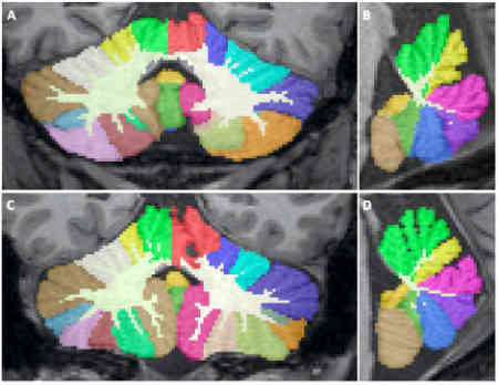
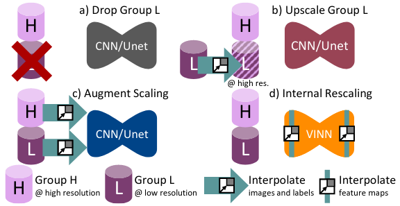
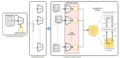
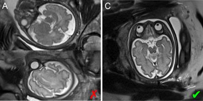
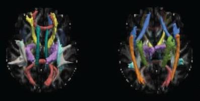
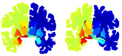
Faber J*, Kügler D*, Bahrami E*, Heinz LS, Timmann D, Ernst TM, Deike-Hofmann K, Klockgether T, van de Warrenburg B, van Gaalen J, Reetz K, Romanzetti S, Oz G, Joers JM, Diedrichsen J, ESMMR Study Group, Reuter M. (*co-first). CerebNet: A fast and reliable deep-learning pipeline for detailed cerebellum sub-segmentation. NeuroImage. 2022.

Key Figure


Henschel L, Kügler D, Reuter M.. Identifying and Combating Bias in Segmentation Networks by leveraging multiple resolutions. Medical Image Computing and Computer-Assisted Interventions, International Conference of (MICCAI). 2022.

Key Figure


Saikat R*, Kügler D*, Reuter M. (*co-first). Are 2.5D approaches superior to 3D deep networks in whole brain segmentation?. Medical Imaging with Deep Learning, Conference (MIDL). 2022.

Key Figure


Henschel L*, Kügler D*, Reuter M. (*co-first). FastSurferVINN: Building Resolution-Independence into Deep Learning Segmentation Methods - A Solution for HighRes Brain MRI. NeuroImage. 2022.

Key Figure


Mauschitz MM, Lohner V, Koch A, Stöcker T, Reuter M, Holz FG, Finger RP, Breteler MMB. Retinal layer assessments as potential biomarkers for brain atrophy in the Rhineland Study. Scientific Reports. 2022.


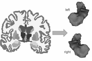
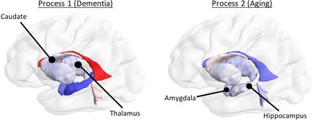
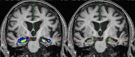
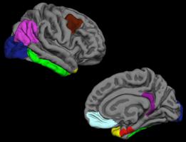
Harms A, Bauer T, Fischbach L, David B, Ernst L, Witt J-A, Diers K, Baumgartner T, Weber B, Radbruch A, Becker AJ, Helmstaedter C, Reuter M, Elger CE, Surges R, Rüber T. Shape description and volumetry of hippocampus and amygdala in temporal lobe epilepsy – A beneficial combination with a clinical perspective. Epilepsy & Behavior. 2022.

Key Figure


Lu R, Aziz NA, Reuter M, Stöcker T, Breteler MMB. Evaluation of the Neuroanatomical Basis of Olfactory Dysfunction in the General Population. JAMA Otolaryngol Head Neck Surg. 2021.

Key Figure


Estrada S, Lu R, Diers K, Zeng W, Ehses P, Stöcker T, Breteler MMB, Reuter M. Automated olfactory bulb segmentation on high resolutional T2-Weighted MRI. NeuroImage. 2021.

Key Figure


Hoffmann M, Turk EA, Gagoski B, Morgan L, Wighton P, Tisdall MD, Reuter M, Adalsteinsson E, Grant PE, Wald LL, van der Kouwe AJW. Rapid head-pose detection for automated slice prescription of fetal-brain MRI. IMA. 2021.

Key Figure


Lu R, Aziz A, Diers K, Stöcker T, Reuter M, Breteler MMB. Insulin resistance accounts for metabolic syndrome-related alterations in brain structure. Human Brain Mapping. 2021.

Key Figure


Henschel L, Reuter M. Parameter Space CNN for Cortical Surface Segmentation. Bildverarbeitung für die Medizin 2020. Informatik aktuell. 2020.

Key Figure


Ewert C, Kügler D, Yendiki A, Reuter M. Learning Anatomical Segmentations for Tractography from Diffusion MRI. MICCAI Workshop on Computational Diffusion MRI. 2020.

Key Figure


Henschel L, Conjeti S, Estrada S, Diers K, Fischl B, Reuter M. FastSurfer - A fast and accurate deep learning based neuroimaging pipeline. NeuroImage. 2020.

Key Figure


Estrada S, Lu R, Conjeti S, Orozco‐Ruiz X, Panos‐Willuhn J, Breteler MMB, Reuter M. FatSegNet: A Fully Automated Deep Learning Pipeline for Adipose Tissue Segmentation on Abdominal Dixon MRI. MRM. 2019.

Key Figure


Estrada S, Conjeti S, Ahmad M, Nassir N, Reuter M. Competition vs. Concatenation in Skip Connections of Fully Convolutional Networks. MICCAI - International Workshop on Machine Learning in Medical Imaging. 2018.

Key Figure


Ahmad M, Conjeti S, Estrada S, Ehses P, Stöcker T, Reuter M. Complex Fully Convolutional Neural Networks for MR Image Reconstruction. MICCAI - International Workshop on Machine Learning for Medical Image Reconstruction. 2018.

Key Figure


Zaretskaya N, Fischl B, Reuter M, Renvall V, Polimeni JR. Advantages of cortical surface reconstruction using submillimeter 7T MEMPRAGE. NeuroImage. 2018.

Key Figure


Schwarz JM, Pracht ED, Brenner D, Reuter M, Stöcker T. GRAPPA reconstructed wave‐CAIPI MP‐RAGE at 7 Tesla. MRM. 2018.

Key Figure


Wachinger C, Nho K, Saykin AJ, Reuter M, Rieckmann A. A Longitudinal Imaging Genetics Study of Neuroanatomical Asymmetry in Alzheimer’s Disease. Biol Psychiatry. 2018.

Key Figure


Ranjan C, Paynabar K, Reuter M, Jafari-Khouzani K. Longitudinal MRI Data Analysis in Presence of Measurement Error but Absence of Replicates. IISE Trans Healthc Syst Eng. 2018.


Wachinger C, Reuter M, Klein T. DeepNAT: Deep convolutional neural network for segmenting neuroanatomy. NeuroImage. 2018.

Key Figure


Wachinger C, Rieckmann A, Reuter M. Latent Processes Governing Neuroanatomical Change in Aging and Dementia. MICCAI. 2017.

Key Figure


Berron D, Schütze H, Cardenas-Blanco A, Fliessbach K, Wagner M, Spottke A, Reuter M, Teipel SJ, Buerger K, Schneider A, Peters O, Nestor P, Priller J, Wiltfang J, Laske C, Jessen F, Düzel E. Object and Scene Memory are Differentially Associated with CSF Markers OF Alzheimer’s Disease and MRI Volumetry. Alzheimers Dement. 2017.


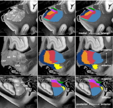
Saygin, ZM, Kliemann D, Iglesias JE, van der Kouwe AJW, Boyd E, Reuter M, Stevens A, Van Leemput K, McKee A, Frosch MP, Fischl B, Augustinack JC. High-resolution magnetic resonance imaging reveals nuclei of the human amygdala: manual segmentation to automatic atlas. NeuroImage. 2017.

Key Figure


Klein A, Ghosh SS, Bao FS, Giard J, Häme Y, Stavsky E, Lee N, Rossa B, Reuter M, Chaibub Neto E, Keshavan A. Mindboggling morphometry of human brains. Comput Biol. 2017.

Key Figure


Aganj I, Iglesias JE, Reuter M, Sabuncu MR, Fischl B. Mid-space-independent deformable image registration. NeuroImage. 2017.

Key Figure


Wachinger C, Salat DH, Weiner M, Reuter M. Whole-brain analysis reveals increased neuroanatomical asymmetries in dementia for hippocampus and amygdala. Brain. 2016.

Key Figure


Ge T, Reuter M, Winkler AM, Holmes AJ, Lee PH, Tirrell LS, Roffman JL, Buckner RL, Smoller JW, Sabuncu MR. Multidimensional heritability analysis of neuroanatomical shape. Nat Commun. 2016.

Key Figure


Iglesias JE, Van Leemput K, Augustinack J, Insausti R, Fischl B, Reuter M. Bayesian longitudinal segmentation of hippocampal substructures in brain MRI using subject-specific atlases. NeuroImage. 2016.

Key Figure


Wachinger C, Reuter M. Domain adaptation for Alzheimer’s disease diagnostics. NeuroImage. 2016.

Key Figure


Yendiki A, Reuter M, Wilkens P, Rosas HD, Fischl B. Joint reconstruction of white-matter pathways from longitudinal diffusion MRI data with anatomical priors. NeuroImage. 2016.

Key Figure


Tisdall MD, Reuter M, Qureshi A, Buckner RL, Fischl B, van der Kouwe A. Prospective motion correction with volumetric navigators (vNavs) reduces the bias and variance in brain morphometry induced by subject motion. NeuroImage. 2016.

Key Figure


Biller A, Reuter M, Patenaude B, Homola GA, Breuer F, Bendszus M, Bartsch AJ. Responses of the Human Brain to Mild Dehydration and Rehydration Explored In Vivo by 1H-MR Imaging and Spectroscopy. Am J Neuroradiol. 2015.

Key Figure


Aganj I, Iglesias JE, Reuter M, Sabuncu MR, Fischl B. Mid-Space-Independent Symmetric Data Term for Pairwise Deformable Image Registration. MICCAI. 2015.

Key Figure


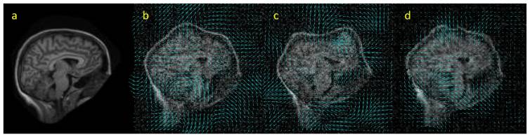
Cash DM, Frost C, Iheme LO, Ünay D, Kandemir M, Fripp J, Salvado O, Bourgeat P, Reuter M, Fischl B, Lorenzi M, Frisoni GB, Pennec X, Pierson RK, Gunter JL, ML Senjem, Jack CR, Guizard JN, Fonov VS, Collins DL, Modat M, Cardoso MJ, Leung KK, Wang H, Das SR, Yushkevich PA, Malone IB, Fox NC, Schott JM, Ourselin S. Assessing atrophy measurement techniques in dementia: Results from the MIRIAD atrophy challenge. NeuroImage. 2015.

Key Figure


Wachinger C, Golland P, Kremen W, Fischl B, Reuter M. BrainPrint: a discriminative characterization of brain morphology. NeuroImage. 2015.

Key Figure


Bron EE, Smits M, van der Flier WM, Vrenken H, Barkhof F, Scheltens P, Papma JM, Steketee RM, Méndez Orellana C, Meijboom R, Pinto M, Meireles JR, Garrett C, Bastos-Leite AJ, Abdulkadir A, Ronneberger O, Amoroso N, Bellotti R, Cárdenas-Peña D, Álvarez-Meza AM, Dolph CV, Iftekharuddin KM, Eskildsen SF, Coupé P, Fonov VS, Franke K, Gaser C, Ledig C, Guerrero R, Tong T, Gray KR, Moradi E, Tohka J, Routier A, Durrleman S, Sarica A, Di Fatta G, Sensi F, Chincarini A, Smith GM, Stoyanov ZV, Sørensen L, Nielsen M, Tangaro S, Inglese P, Wachinger C, Reuter M, van Swieten JC, Niessen WJ, Klein S. Standardized evaluation of algorithms for computer-aided diagnosis of dementia based on structural MRI: The CADDementia challenge. NeuroImage. 2015.

Key Figure


Reuter M, Tisdall MD, Qureshi A, Buckner RL, van der Kouwe AJW, Fischl B. Head motion during MRI acquisition reduces gray matter volume and thickness estimates. NeuroImage. 2015.

Key Figure


Wachinger C, Golland P, Magnain C, Fischl B, Reuter M. Multi-Modal Robust Inverse-Consistent Linear Registration. Hum Brain Mapp. 2015.

Key Figure


Aganj I, Reuter M, Sabuncu MR, Fischl B. Avoiding symmetry-breaking spatial non-uniformity in deformable image registration via a quasi-volume-preserving constraint. NeuroImage. 2015.

Key Figure


Wachinger C, Golland P, Reuter M. BrainPrint : Identifying Subjects by Their Brain. MICCAI. 2014.

Key Figure


Wachinger C, Batmanghelich K, Golland P, Reuter M. BrainPrint in the Computer-Aided Diagnosis of Alzheimer’s Disease. MICCAI Challenge on Computer-Aided Diagnosis of Dementia Based on Structural MRI Data. 2014.

Key Figure


Wachinger C, Golland P, Reuter M, Wells W. Gaussian process interpolation for uncertainty estimation in image registration. MICCAI. 2014.

Key Figure


Wade BS, Joshi SH, Reuter M, Blumenthal JD, Toga AW, Thompson PM, Giedd JN. Effects of sex chromosome dosage on corpus callosum morphology in supernumerary sex chromosome aneuploidies. Biol Sex Differ. 2014.

Key Figure


Wang H, Zhu J, Reuter M, Vinke LN, Yendiki A, Boas DA, Fischl B, Akkin T. Cross-validation of serial optical coherence scanning and diffusion tensor imaging: a study on neural fiber maps in human medulla oblongata. NeuroImage. 2014.

Key Figure


Sabuncu MR, Bernal-Rusiel JL, Reuter M, Greve DN, Fischl B. Event time analysis of longitudinal neuroimage data. NeuroImage. 2014.

Key Figure


Reuter M, Gerstner ER, Rapalino O, Batchelor TT, Rosen B, Fischl B. Impact of MRI head placement on glioma response assessment. J Neurooncol. 2014.

Key Figure


Augustinack JC, Magnain C, Reuter M, van der Kouwe AJW, Boas D, Fischl B. MRI Parcellation of Ex Vivo Medial Temporal Lobe. NeuroImage. 2014.

Key Figure


Andronesi OC, Bhat H, Reuter M, Mukherjee S, Caravan P, Rosen BR. Whole brain mapping of water pools and molecular dynamics with rotating frame MR relaxation using gradient modulated low-power adiabatic pulses. NeuroImage. 2014.

Key Figure


Fujimoto K, Polimeni JR, van der Kouwe AJW, Reuter M, Kober T, Benner T, Fischl B, Wald LL. Quantitative comparison of cortical surface reconstructions from MP2RAGE and multi-echo MPRAGE data at 3 and 7 T. NeuroImage. 2014.

Key Figure


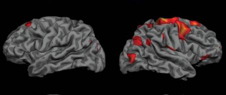
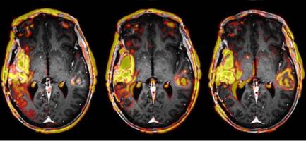
Adriaanse SM, Dijk KRA, Ossenkoppele R, Reuter M, Tolboom N, Zwan MD, Yaqub M, Boellaard R, Windhorst AD, Flier WM, Scheltens, P, Lammertsma AA, Barkhof F, Berckel BNM. The effect of amyloid pathology and glucose metabolism on cortical volume loss over time in Alzheimer’s disease. European Journal of Nuclear Medicine and Molecular Imaging. 2014.

Key Figure


Rosas HD, Doros G, Gevorkian S, Malarick K, Reuter M, Coutu JP, Triggs T, Wilkens P, Matson W, Salat DH, Hersch SM. PRECREST: A phase II prevention and biomarker trial of creatine in at-risk Huntington disease. Neurology. 2014.

Key Figure


Magnain C, Augustinack JC, Reuter M, Wachinger C, Frosch MP, Ragan T, Akkin T, Wedeen VJ, Boas DA, Fischl B. Blockface histology with optical coherence tomography: A comparison with Nissl staining. NeuroImage. 2014.

Key Figure


Bernal-Rusiel JL, Reuter M, Greve DN, Fischl B, Sabuncu MR. Spatiotemporal Linear Mixed Effects Modeling for the Mass-univariate Analysis of Longitudinal Neuroimage Data. NeuroImage. 2013.

Key Figure


Aganj I, Reuter M, Sabuncu MR, Fischl B. Symmetric non-rigid image registration via an adaptive quasi-volume-preserving constraint. ISBI. 2013.

Key Figure


Bernal-Rusiel JL, Greve DN, Reuter M, Fischl B, Sabuncu MR. Statistical analysis of longitudinal neuroimage data with Linear Mixed Effects models. NeuroImage. 2013.


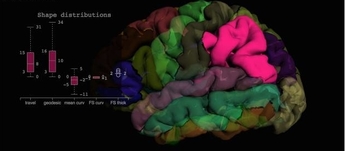
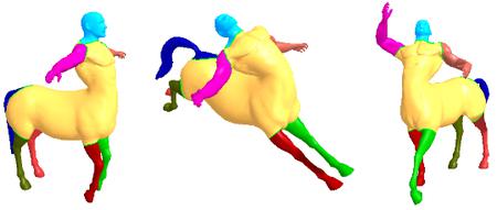
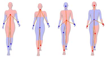
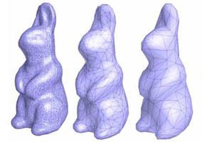
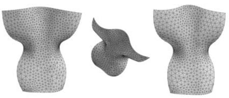
Lian Z, Godil A, Bustos B, Daoudi M, Hermans J, Kawamura S, Kurita Y, Lavoué G, Van Nguyen H, Ohbuchi R, Ohkita Y, Ohishi Y, Porikli F, Reuter M, Sipiran I, Smeets D, Suetens P, Tabia H, Vandermeulen D. A comparison of methods for non-rigid 3D shape retrieval. Pattern Recognition. 2013.

Key Figure


Tisdall MD, Hess AT, Reuter M, and Meintjes EM, Fischl B, van der Kouwe AJW. Volumetric navigators for prospective motion correction and selective reacquisition in neuroanatomical MRI. Magnetic Resonance in Medicine. 2012.

Key Figure


Adriaanse S, van Dijk K, Ossenkoppele R, Reuter M, Tolboom N, Zwan M, Yaqub M, Boellaard R, Windhorst A, Van der Flier W, Scheltens, P, Barkhof F, Lammertsma A, van Berckel B. Exploring the biomarker cascade model: Relating cortical volume loss to [11C]PIB, [18F]FDG and MMSE over time in Alzheimer’s disease patients and normal controls. Alzheimer’s & Dementia. 2012.


Reuter M, Schmansky NJ, Rosas HD, Fischl B. Within-Subject Template Estimation for Unbiased Longitudinal Image Analysis. NeuroImage. 2012.

Key Figure


Rosas HD, Reuter M, Doros G, Lee SY, Triggs T, Malarick K, Fischl B, Salat DH, Hersch SM. A tale of two factors: What determines the rate of progression in Huntington’s disease? A longitudinal MRI study. Movement Disorders. 2011.

Key Figure


Sabuncu MR, Desikan RS, Sepulcre J, Yeo BTT, Liu H, Schmansky NJ, Reuter M, Weiner MW, Buckner RL, Sperling RA, Fischl B, Alzheimer’s Disease Neuroimaging Initiative. The Dynamics of Cortical and Hippocampal Atrophy in Alzheimer Disease. Arch Neurol.. 2011.

Key Figure


Reuter M, Fischl B. Avoiding Asymmetry-Induced Bias in Longitudinal Image Processing. NeuroImage. 2011.


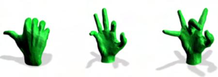
Lian Z, Godil A, Bustos B, Daoudi M, Hermans J, Kawamura S, Kurita Y, Lavoué G, Nguyen HV, Ohbuchi R, Ohkita Y, Ohishi Y, Porikli F, Reuter M, Sipiran I, Smeets D, Suetens P, Tabia H, Vandermeulen D. SHREC’11 track: shape retrieval on non-rigid 3D watertight meshes. Eurographics - 3DOR’11 Workshop. 2011.

Key Figure


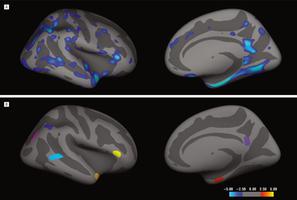
Desikan RS, Sabuncu MR, Schmansky NJ, Reuter M, Cabral HJ, Hess CP, Weiner MW, Biffi A, Anderson CD, Rosand J, Salat DH, Kemper TL, Dale AM, Sperling RA, Fischl B, Alzheimer’s Disease Neuroimaging Initiative. Seletive Disruption of the Cerebral Neocortex in Alzheimer’s Disease. PLoS ONE. 2010.

Key Figure


Reuter M, Rosas HD, Fischl B. Highly Accurate Inverse Consistent Registration: A Robust Approach. NeuroImage. 2010.

Key Figure


Reuter M. Hierarchical Shape Segmentation and Registration via Topological Features of Laplace-Beltrami Eigenfunctions. International Journal of Computer Vision. 2010.

Key Figure


Reuter M, Biasotti S, Giorgi D, Patanè G, Spagnuolo M. Discrete Laplace-Beltrami operators for shape analysis and segmentation. Computers & Graphics. 2009.

Key Figure


Reuter M, Wolter FE, Shenton M, Niethammer M. Laplace-Beltrami Eigenvalues and Topological Features of Eigenfunctions for Statistical Shape Analysis. Computer-Aided Design. 2009.

Key Figure


Reuter M, Mikkelsen TS, Sherbrooke EC, Maekawa T, Patrikalakis NM. Solving nonlinear polynomial systems in the barycentric Bernstein basis. The Visual Computer. 2008.

Key Figure


Reuter M. Can one hear Shape?. GAMM07 and ICIAM07. 2007.

Key Figure


Reuter M, Niethammer M, Wolter FE, Bouix S, Shenton M. Global Medical Shape Analysis Using the Volumetric Laplace Spectrum. CW. 2007.

Key Figure


Niethammer M, Reuter M, Wolter FE, Bouix S, Peinecke N, Koo MS, Shenton M. Global Medical Shape Analysis using the Laplace-Beltrami Spectrum. MICCAI. 2007.

Key Figure


Wolter FE, Reuter M, Peinecke N. Geometric Modeling for Engineering Applications. Encyclopedia of Computational Mechanics. 2007.

Key Figure


Peinecke N, Wolter FE, Reuter M. Laplace Spectra as Fingerprints for Image Recognition. Computer-Aided Design. 2007.

Key Figure


Reuter M. Laplace Spectra for Shape Recognition. BoD. 2006.

Key Figure


Reuter M, Wolter FE, Peinecke N. Laplace-Beltrami spectra as “Shape-DNA” of surfaces and solids. Computer-Aided Design. 2006.

Key Figure


Reuter M, Wolter FE, Peinecke N. Laplace-Spectra as Fingerprints for Shape Matching. SPM. 2005.

Key Figure


Wolter FE, Peinecke N, Reuter M. Geometric Modeling of Complex Shapes and Engineering Artifacts. Encyclopedia of Computational Mechanics. 2004.

Key Figure