We develop open, validated computational tools at the intersection of deep learning, medical image computing, and computational geometry.
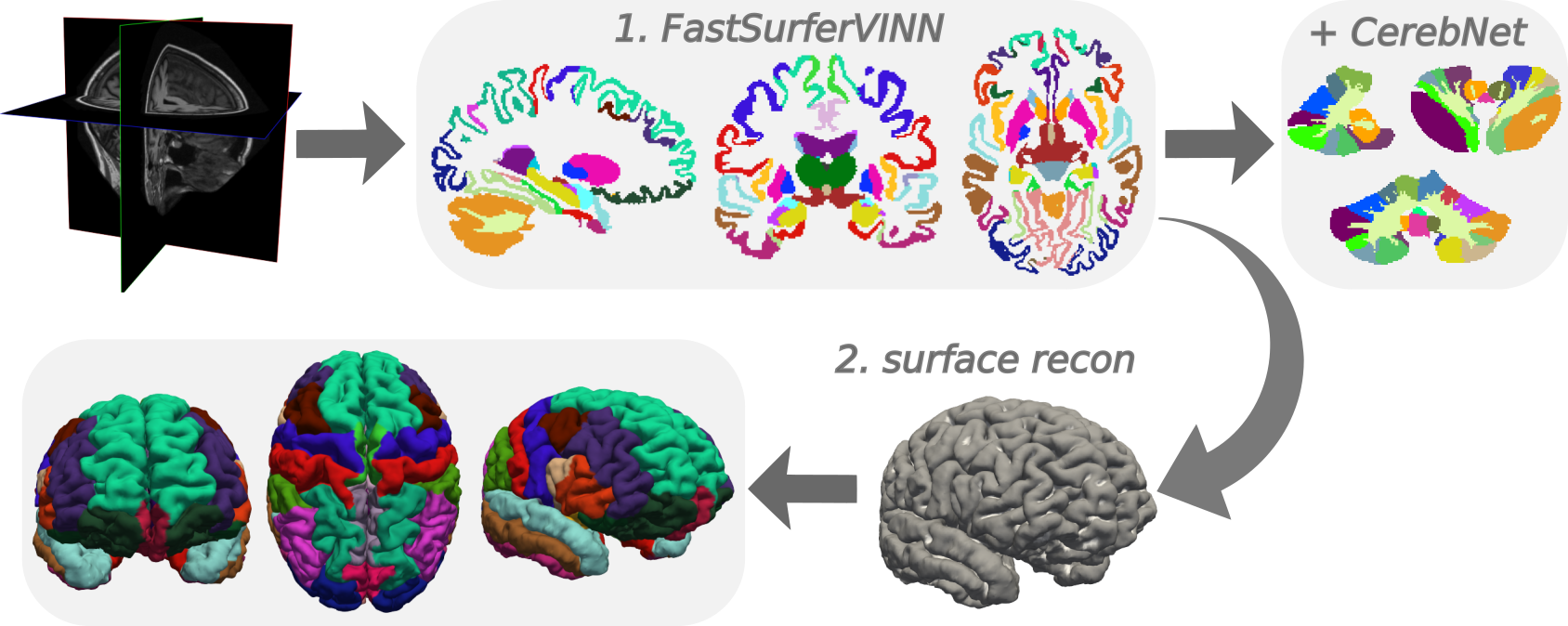
Fast, accurate, deep-learning pipeline for whole-brain segmentation in under a minute and full surface reconstruction in about an hour. Produces FreeSurfer-conform outputs for large-cohort and clinical use.
 deep-mi/FastSurfer
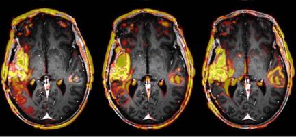
GPU-accelerated Python toolkit for neuroimaging registration — same-modality, cross-modal, boundary-based, and multi-timepoint — plus transform and volume utilities. Replaces FreeSurfer's mri_robust_register, mri_coreg, and bbregister. Usable from the command line or as a library.
deep-mi/neuroreg
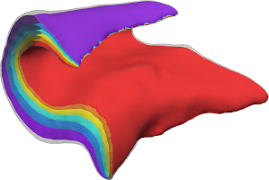
Automated geometry-based method for hippocampal shape and thickness analysis from structural MRI, enabling fine-grained morphometric studies of the hippocampus.
deep-mi/hipsta
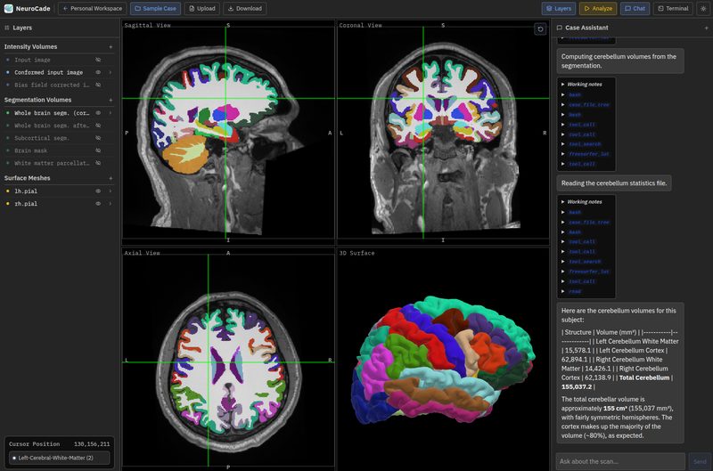
AI-powered neuroimaging platform combining an MRI viewer with integrated pipelines like FastSurfer. Run complex structural imaging workflows through a GUI or conversational AI — no command line required.
deep-mi/NeuroCade
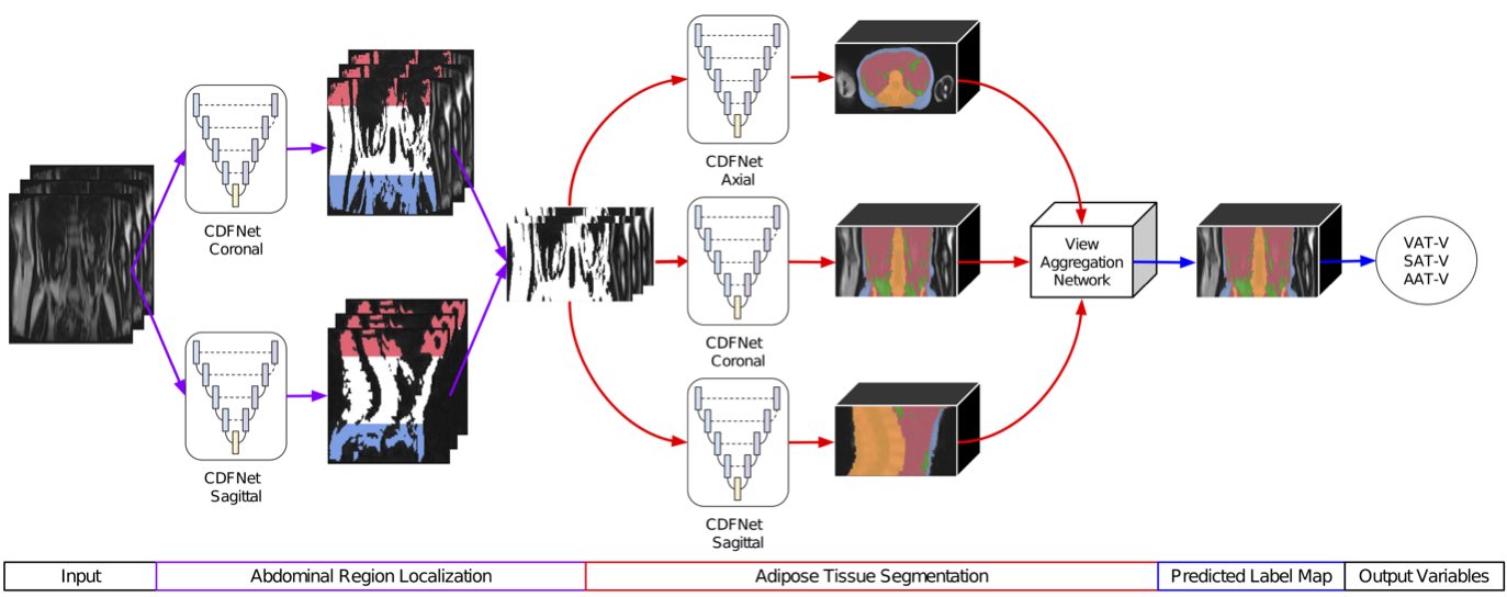
Automated adipose tissue segmentation and field-of-view estimation in whole-body Dixon MRI, enabling large-scale body composition studies.
deep-mi/FatSegNet
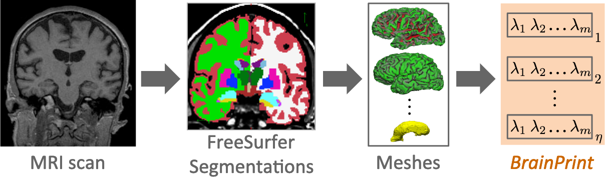
Shape and asymmetry analysis of neuroanatomical structures using Laplace-Beltrami spectral descriptors for group studies and individual subject classification.
Deep-MI/BrainPrint
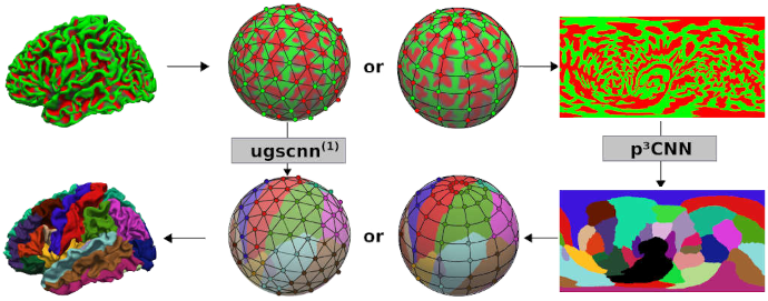
Cortical surface parcellation using projection-based 3D convolutional neural networks operating on spherical mesh representations.
Learn more →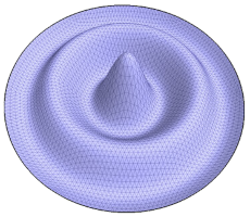
Python library for geometric mesh processing and finite element method (FEM) differential geometry computations on triangular and tetrahedral meshes.
deep-mi/LaPy
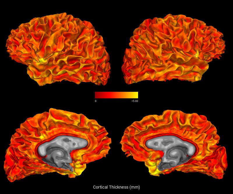
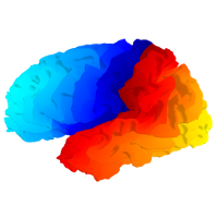
Python/OpenGL tool for rendering surface meshes with color overlays and generating publication-quality screenshots — fully offscreen (no GPU required), runs in Docker and headless environments, as well as from the command line, Jupyter notebooks, or an interactive desktop GUI.
deep-mi/WhipperSnapPy
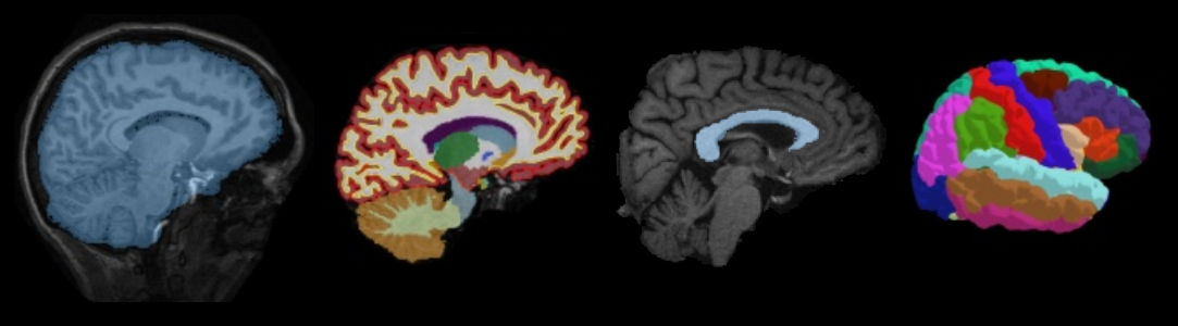
Automated quality check tools for surface-based morphometry outputs from FastSurfer and FreeSurfer, enabling quality control at scale.
deep-mi/qatools-python
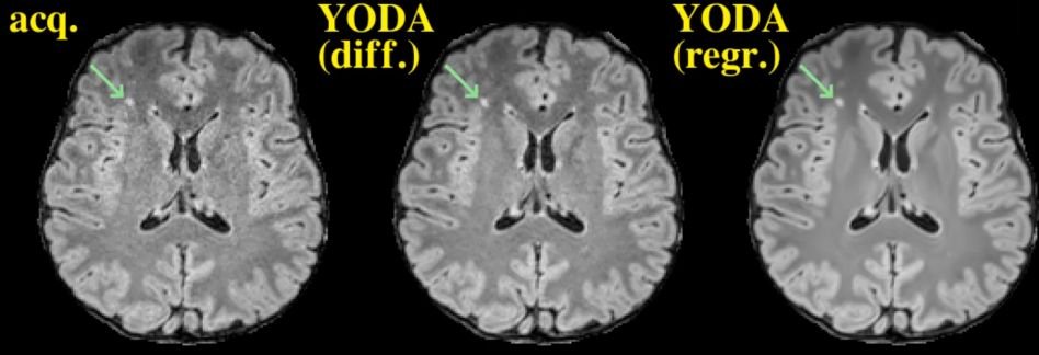
State-of-the-Art framework for medical image translation (e.g. FLAIR generation) optimizing for information fidelity rather than perceptual realism (and thus noise replication).
deep-mi/YODA
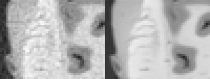
MRI denoiser, that actually (!) removes physical acquisition noise, squeezing every bit of information out of (already-decent) MR images.
deep-mi/YADO
All code is open source and available on github.com/Deep-MI.
Our work spans computational neuroimaging, geometric deep learning, and medical image analysis. In close collaboration with clinical and industrial partners, we develop next-generation methods for large-scale biomedical imaging datasets.
Deep-learning methods for reliable image segmentation, registration, and reconstruction — with a focus on sensitivity in longitudinal studies and generalizability across scanners and protocols.
Statistical modeling and computer vision for extracting clinically relevant biomarkers from large datasets — supporting computer-aided diagnosis, prognosis, and personalized medicine.
Differential and computational geometry methods for shape analysis, spectral descriptors, and surface-based morphometry of neuroanatomical structures.
Understanding brain development and neurodegeneration (aging, Alzheimer's, Huntington's disease) via multi-modal imaging of large population cohorts such as the Rhineland Study.
All research from the Deep-MI lab is backed by peer-reviewed publications. You can find the full list on the publications page or on Google Scholar.
 Deep-MI
Lab
Deep-MI
Lab