BrainPrint - Neuroanatomical Shape Analysis

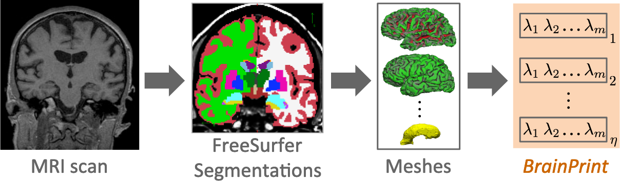
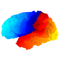
Our BrainPrint tools provide shape descriptors of neuroanatomical structures and require a FreeSurfer or FastSurfer segmentation as a pre-processing step. BrainPrint is based on “ShapeDNA” a spectral shape descriptor that is well suited for the analysis of non-rigid bendable shapes like biological structures.

Spectral Shape Analysis

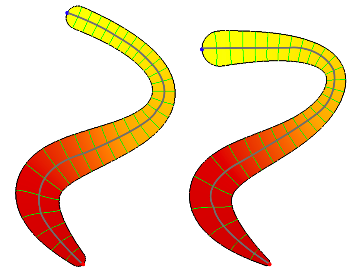
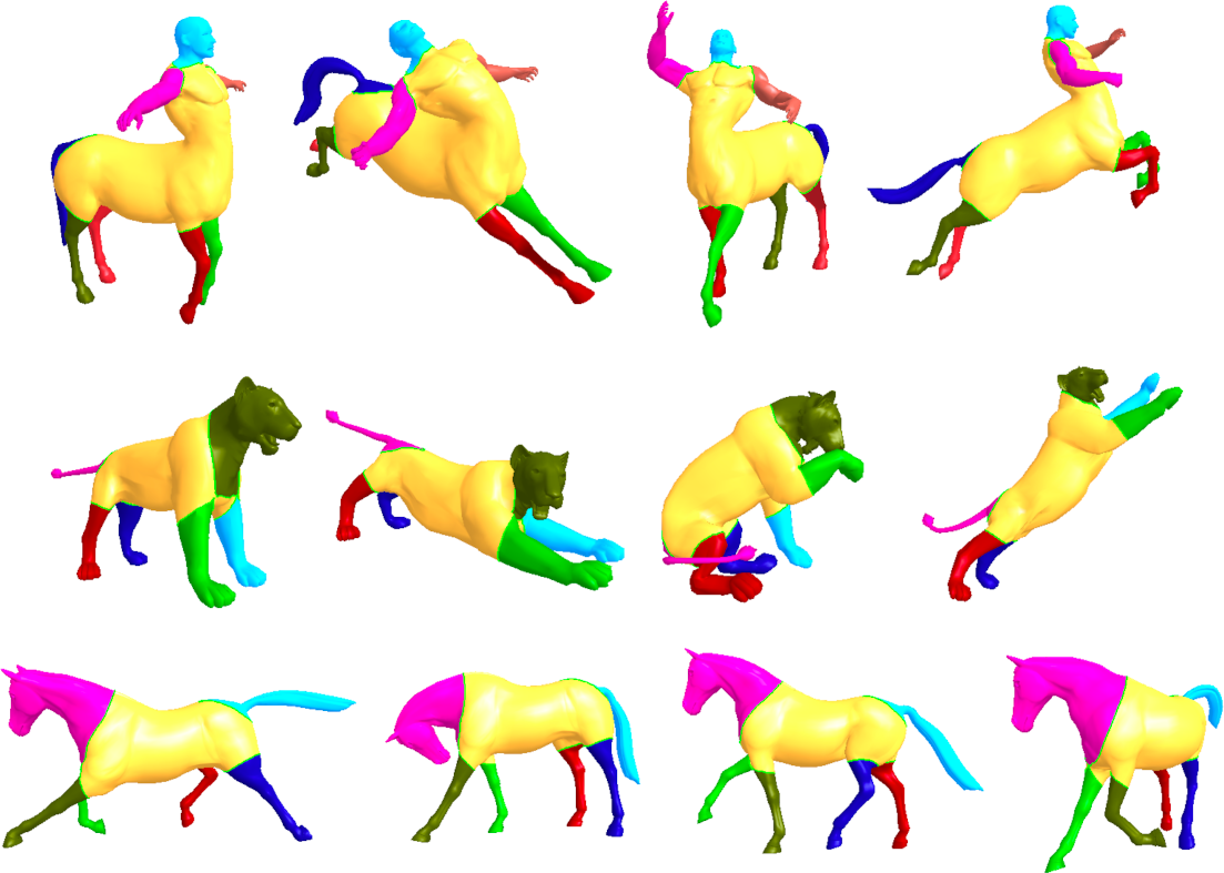
In spectral shape analysis we employ the spectrum of the Laplace-Beltrami operator as a shape descriptor for the analysis of shape differences. The main advantage is that this descriptor is isometry invariant. Isometry invariance means that distances measured along the surface stay the same. So a hand with different finger positions or a person in different body postures will be (near) isometric, as not much streching is involved. The distance from the nose to the foot is fixed for different body postures, if measured along the surface (as opposed to measuring it in the embedding space). Thus we are able to identify similar deformable objects even if they cannot be aligned/compared with a rigid transformation!

Eigenmode

Fig 1. First non-constant eigenmode for similar shapes. Red and blue dots at the tips denote the extrema, the green curves are some level sets. The Reeb graph (gray curve) approximates the medial axis.

Registration

Fig 2. Registration of segments across near isometric shapes. Same color indicates registered parts across poses of the same model.

BrainPrint

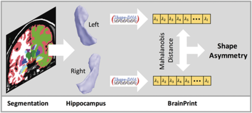
We have extended this work to analyze brain shape changes (with respect to symmetry, heritability, computer-aided diagnosis of neurodegenerative disease, etc). The code for the BrainPrint tools is available on github. Documentation and API can be found here.

BrainPrint

Fig 3. Overview of the computation of the BrainPrint. First, MRI scans are processed with FreeSurfer to obtain segmentations of cortical and subcortical structures. Second, a mesh is created for each brain structure. Third, the ShapeDNA is computed for all meshes, constituting the BrainPrint.

Asymmetry

Fig 4. Based on a brain segmentation, we create meshes from lateralized structures, e.g. the hippocampus. The computation of the shape descriptor ShapeDNA yields the characteristic spectrum of the shape, which forms the BrainPrint. The Mahalanobis distance between the spectra of both hemispheres results in the shape asymmetry.